Artificial intelligence in breast imaging has moved beyond theoretical validation and controlled studies into day-to-day clinical use. The challenge now is not whether AI works in principle, but how it performs within real-world workflows, across diverse patient populations, imaging systems, and service pressures.
In practice, implementation rarely follows a linear path from development to deployment. Instead, it is shaped by local infrastructure, clinician confidence, regulatory requirements, and the realities of existing screening and diagnostic pathways.
From Standalone Tools to Embedded Systems
Early AI solutions were often introduced as separate platforms—reviewed alongside existing PACS systems or used in parallel with radiologist interpretation. This model quickly revealed limitations: duplication of effort, workflow disruption, and inconsistent adoption.
The current direction is integration. AI is increasingly embedded directly into imaging workflows, supporting tasks such as:
- Prioritising abnormal studies for faster review
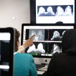
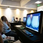
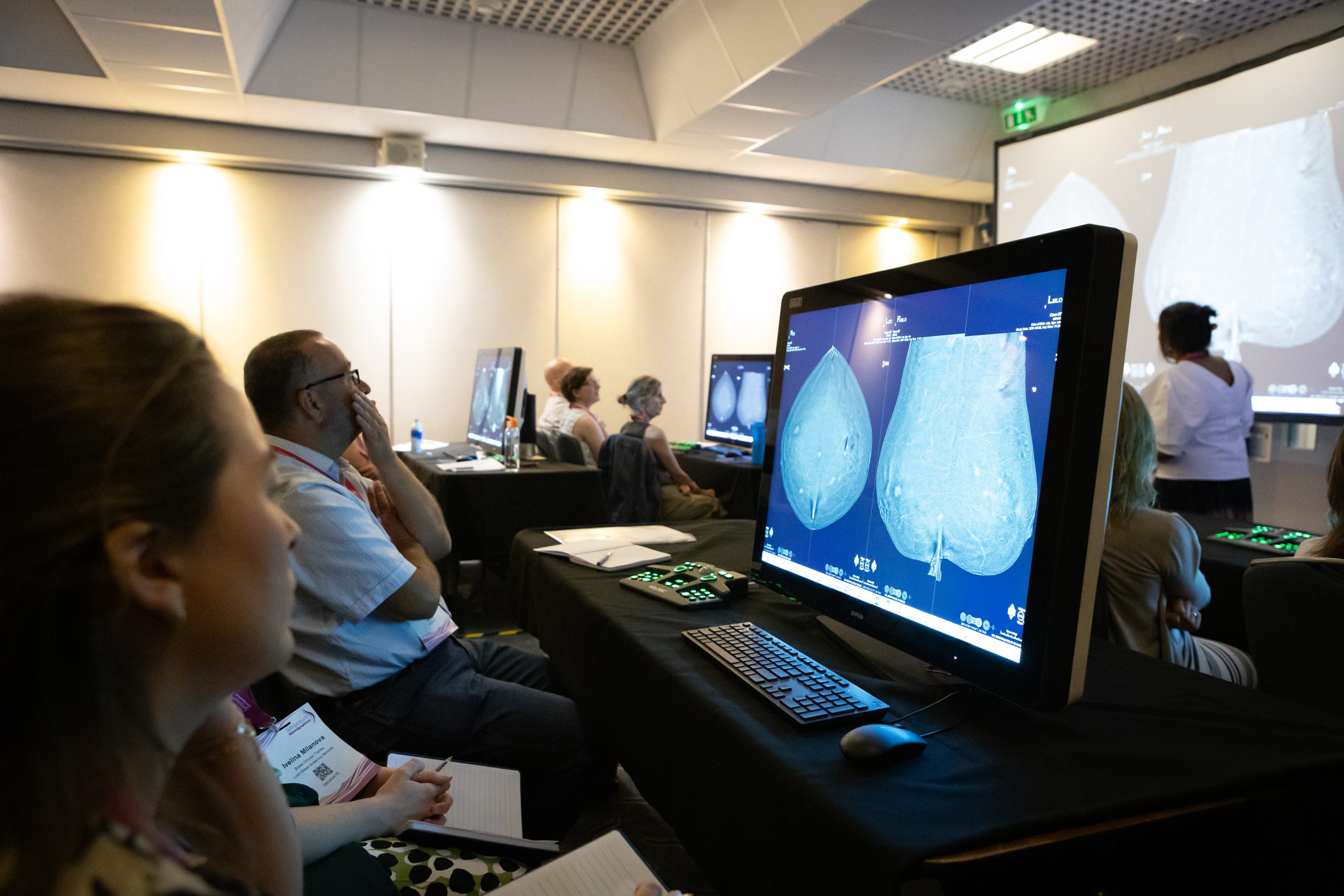
- Highlighting regions of interest on mammograms
- Assisting with breast density classification
- Supporting structured reporting and documentation
When AI becomes part of the native workflow, rather than an external step, adoption becomes more sustainable and clinically meaningful.
The Importance of Workflow Fit
Successful implementation depends less on algorithm performance alone and more on how well the system fits into clinical practice.
Key considerations include:
- Time added or saved per case
- Alignment with radiologist reading patterns
- Integration with reporting systems
- Clarity of AI outputs and explanations
Even highly accurate tools can fail in practice if they increase cognitive load or disrupt established routines. Conversely, modest gains in accuracy may be highly valuable if they significantly improve efficiency or consistency.
Real-World Performance vs Controlled Studies
A recurring theme in deployment is the difference between controlled evaluation environments and real-world conditions.
In clinical practice, AI must contend with:
- Variability in image quality
- Differences in equipment across sites
- Diverse patient demographics
- Incomplete or noisy clinical data
This variability often exposes performance gaps not visible in curated datasets. As a result, ongoing monitoring and recalibration are becoming essential parts of implementation strategies.
Human-AI Collaboration in Practice
Rather than replacing radiologists, AI is increasingly positioned as a collaborative tool. The most effective implementations support a “human-in-the-loop” model, where:
- AI assists with detection and prioritisation
- Clinicians retain final interpretation and responsibility
- Discrepancies are reviewed and used for system learning
This collaboration is not static. Over time, it reshapes reading patterns, training needs, and even multidisciplinary discussion structures.
Barriers to Adoption
Despite growing clinical evidence, several barriers remain:
- Integration complexity across legacy systems
- Variation in regulatory approval and reimbursement pathways
- Limited interoperability between vendors
- Concerns around liability and accountability
- Need for robust validation in local populations
These challenges mean that adoption is often incremental rather than uniform, even within advanced healthcare systems.
Conclusion
Real-world AI implementation in breast imaging is less about deployment of a single technology and more about redesigning how imaging services operate. The shift from protocol to practice requires alignment between algorithms, clinicians, and clinical systems.
Success depends not only on what AI can do in isolation, but on how effectively it supports decision-making within the realities of everyday care.